
The field of neuroscience has recently witnessed a groundbreaking development in the form of a new two-photon fluorescence microscope that has the capability to capture high-speed images of neural activity at cellular resolution. This innovation is poised to revolutionize the way researchers study neural networks in real time, shedding light on critical aspects of brain function and neurological diseases.
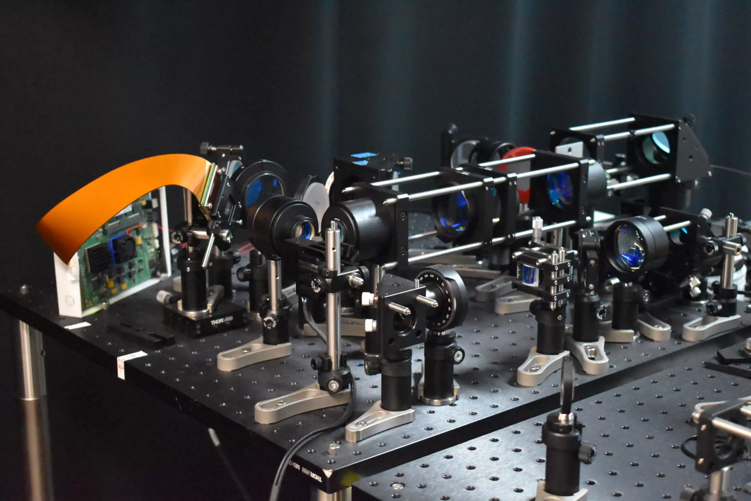
Unlike traditional two-photon microscopy techniques, the new microscope boasts an adaptive sampling scheme and utilizes line illumination instead of point illumination. This novel approach enables researchers to image neuronal activity in a mouse cortex at speeds ten times faster than conventional methods while also reducing the potential harm to brain tissue significantly.
The implications of this advancement are profound, with the potential to deepen our understanding of fundamental brain functions such as learning, memory, and decision-making. By providing a tool for observing neuronal activity in real time, researchers can now explore the dynamics of neural networks with unprecedented clarity, offering valuable insights into the pathology of neurological diseases at their earliest stages.
One of the key innovations of this new microscope is the adaptive sampling strategy, which replaces the traditional point illumination with a short line of light. This allows for a larger area of brain tissue to be imaged at once, significantly speeding up the imaging process while minimizing the risk of damage to living tissue. Additionally, the use of a digital micromirror device (DMD) enables precise targeting of active neurons, further enhancing the efficiency and precision of the imaging technique.
The researchers demonstrated the capabilities of the new microscope by imaging calcium signals in living mouse brain tissue at an impressive speed of 198 Hz. This rapid imaging rate opens up possibilities for monitoring quick neuronal events that would be missed by slower methods. Furthermore, the adaptive line-excitation technique coupled with advanced computational algorithms allows for the isolation of individual neuron activity, offering a deeper understanding of complex neural interactions and the functional organization of the brain.
Looking ahead, the researchers are focused on expanding the capabilities of the microscope by integrating voltage imaging for a direct and rapid readout of neural activity. This next step will enable a more comprehensive exploration of neural dynamics and further enhance our understanding of brain function. Additionally, the researchers aim to optimize the usability and size of the microscope to facilitate its integration into a wide range of neuroscience applications.
The new two-photon fluorescence microscope represents a significant leap forward in neural imaging technology, offering unprecedented speed, resolution, and precision in observing neuronal activity. This advancement holds great promise for unraveling the complexities of the brain and advancing our knowledge of neurological disorders, paving the way for new discoveries in neuroscience research.